Long Bone Diagram / Quizlet Anatomy Bones - Download 3,638 diagram bone stock illustrations, vectors & clipart for free or amazingly low rates!
Dapatkan link
Facebook
X
Pinterest
Email
Aplikasi Lainnya
Long Bone Diagram / Quizlet Anatomy Bones - Download 3,638 diagram bone stock illustrations, vectors & clipart for free or amazingly low rates!. Long bone diagram unlabled manual e books. Start studying anatomy bone diagram long bone. The humerus and the femur are corresponding bones of the arms and legs, respectively. Long bones, especially the femur and tibia, are subjected to most of the load during daily activities and they are crucial for skeletal mobility. The inset shows the lamellae of the compacta arranged in osteons, i.e., vascular canals surrounded by concentric layers of bone.
Long bone diagram unlabled manual e books. Bone diagram barca fontanacountryinn com. Bone is found in the shafts of long bone and consists of various cylindrical units named as haversian system 47. Inside this is a layer of spongy (cancellous) bone which contains red bone marrow. Microscopic bone anatomy human body diagram.
Topic 1 from www.gomlc.com The outside of the flat bone consists of a layer of connective tissue called the periosteum. Bone long blood diaphysis vector anatomical anatomy articular biology body calcium cartilage cell compact detail diagram education educational endosteum epiphysis forelimb health healthy human humerus illustration joint long bone marrow medical medicine organ orthopedic periosteum red. When a human finishes growing these parts fuse together. Anatomy bone coloring sheet sketch coloring page. For today we show you some photos of diagram of a long bone and each. The humerus and the femur are corresponding bones of the arms and legs, respectively. 12 photos of the diagram of a long bone. Download scientific diagram | 1 structure and components of long bone.
The outside of the flat bone consists of a layer of connective tissue called the periosteum.
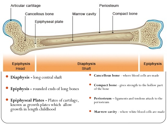
While their parts are similar in general, their structure has been adapted to differing functions. Choose from 500 different sets of flashcards about long bone diagram on quizlet. Human anatomy diagrams show internal organs, cells, systems, conditions, symptoms and sickness information and/or tips for healthy living. Long bones are those that are longer than they are wide. The end of the long bone is the epiphysis and the shaft is the diaphysis. They are one of five types of bones: A typical long bone shows the gross anatomical characteristics of bone. Download scientific diagram | 1 structure and components of long bone. The medullary cavity contains red bone long bones follow the process of endochondral ossification where the diaphysis grows inside of cartilage from a primary ossification center until it. Bone marrow (see diagram below) produces stem cells, such as. Bone diagram barca fontanacountryinn com. Long bones have a spongy bone on their ends but have a hollow medullary cavity in the middle of the diaphysis. The humerus and the femur are corresponding bones of the arms and legs, respectively.
First lets strike out options that we dont need. Choose from 500 different sets of flashcards about long bone diagram on quizlet. Sectional diagram of a long bone. Learn about long bone diagram with free interactive flashcards. When a human finishes growing these parts fuse together.
Human Anatomy Body - Page 2 of 160 - Human Anatomy for ... from www.anatomylibrary99.com First lets strike out options that we dont need. When a human finishes growing these parts fuse together. Find out where this is usually located and, if it is present, label it on your bone. While their parts are similar in general, their structure has been adapted to differing functions. Inside this is a layer of spongy (cancellous) bone which contains red bone marrow. The inset shows the lamellae of the compacta arranged in osteons, i.e., vascular canals surrounded by concentric layers of bone. Download 3,638 diagram bone stock illustrations, vectors & clipart for free or amazingly low rates! 12 photos of the diagram of a long bone.
Choose from 500 different sets of flashcards about long bone diagram on quizlet.
Medical, educational, science poster vector illustrationn. First lets strike out options that we dont need. The other primary skeletal component of. Epiphysis, metaphysis, and diaphysis (see the image diagram depicting tight coupling of osteoblast and osteoclast that allows remodeling to occur. Coronal temporal bone computed tomography image. Bone is found in the shafts of long bone and consists of various cylindrical units named as haversian system 47. Find out where this is usually located and, if it is present, label it on your bone. As shown in figure 2. Download 3,638 diagram bone stock illustrations, vectors & clipart for free or amazingly low rates! Learn about long bone diagram with free interactive flashcards. For today we show you some photos of diagram of a long bone and each. This page is about labeled long bone anatomy diagram,contains labeling a long bone diagram,flashcards anatomy lab practical 1 biology blog, these pictures of this page are about:labeled long bone anatomy diagram. Start studying anatomy bone diagram long bone.
When a human finishes growing these parts fuse together. Choose from 500 different sets of flashcards about long bone diagram on quizlet. Long, short, flat, irregular and sesamoid. For today we show you some photos of diagram of a long bone and each. Long bones, especially the femur and tibia, are subjected to most of the load during daily activities and they are crucial for skeletal mobility.
Long Bone Anatomy | Human Anatomy Quiz - Quizizz from s3-us-west-2.amazonaws.com Each system contains haversian canals surrounded by concentric lamellae of bone tissue 48. Helps keep bones light in weight epiphyseal line line showing where growth plate used to be. Medical, educational, science poster vector illustrationn. Lab 9 overview for lab practical. Choose from 500 different sets of flashcards about long bone diagram on quizlet. Long bone diagram unlabled manual e books. Medivisuals normal foot anatomy medical illustration. Start studying anatomy bone diagram long bone.
The inset shows the lamellae of the compacta arranged in osteons, i.e., vascular canals surrounded by concentric layers of bone.
Bone diagram barca fontanacountryinn com. Medivisuals normal foot anatomy medical illustration. Long bones, especially the femur and tibia, are subjected to most of the load during daily activities and they are crucial for skeletal mobility. The long bones are those that are longer than they are wide. Human anatomy for muscle reproductive and skeleton. The long bones are those that are longer than they are wide. First lets strike out options that we dont need. Choose from 500 different sets of flashcards about long bone diagram on quizlet. Inside this is a layer of spongy (cancellous) bone which contains red bone marrow. They are one of five types of bones: Find out where this is usually located and, if it is present, label it on your bone. Anatomy of a long bone anna s anatomy websit. The outside of the flat bone consists of a layer of connective tissue called the periosteum.
Peaky Blinders Wallpaper 4K : Peaky Blinders Wallpapers - Wallpaper Cave / Download and use 60+ peaky blinders stock photos for free. . View and share our peaky blinders wallpapers post and browse other hot wallpapers, backgrounds and images. In compilation for wallpaper for peaky blinders, we have 27 images. We determined that these pictures can also depict a cast. Also you can download all wallpapers pack with peaky blinders free, you just need click red download button on the right. In this tv show collection we have 27 wallpapers. 1080x2220 peaky blinders season 4 poster 1080x2220 resolution. We determined that these pictures can also depict a cast. Peaky blinders hd wallpapers for desktop download 480 854 peaky. Adorable wallpapers > tv show > peaky blinders wallpapers (31 wallpapers). , peaky blinders wallpaper peaky blinders pinterest wallpapers 1920×1080. Peaky Blinders 4k D...
Banner Ayam Geprek Cdr : desain.ratuseo.com - Warung ayam geprek dahsyat chicken as food logo, chicken, food, animals png. . Download vektor desain kartu ucapan tasyakuran nama anak format coreldraw ( cdr ) disini cari v ektor corel draw gratis lainnya. Banner spanduk toko perlengkapan listrik dan elektronik cdr. By yudha richo post a comment. Find download free graphic resources for ayam. Contoh desain banner spanduk pengajian walimatul ursy cdr. Harga cetak spanduk dihitung dari luas per meter persegi dengan harga per meter rp. Banner spanduk ayam geprek terbaru 2018 cdr banner spanduk. Kalender 2021 lengkap (corel x3 dan x7). Banner ayam geprek cdr / contoh spanduk makanan ringan : Xbanner ayam geprek sewot buat design sesukamu. Banner Ayam Geprek Cdr / Spanduk Ayam Geprek Mudah - Jones ... from lh3.googleusercontent.com Banner spanduk ayam geprek ...
Salmonella Species - Scientists figure out Salmonella bacteria infect plants ... / While some of the infections can be easily treated, some of. . Identification of salmonella species issue no: While some of the infections can be easily treated, some of. Food is the source for most of these illnesses. It is usually characterized by acute onset of fever. Primarily cause enteritis and septicemia; Food is the source for most of these illnesses. It is usually characterized by acute onset of fever. Enterica is the type species and is further divided into six subspecies that include over 2. Identification of salmonella species issue no: Within 2 species, salmonella bongori and salmonella enterica, over 2500 different salmonellosis is a disease caused by the bacteria salmonella. Salmonella- The Most Common Bacterial Foodborne Disease ... from safefoodalliance.com ...


Komentar
Posting Komentar